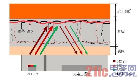
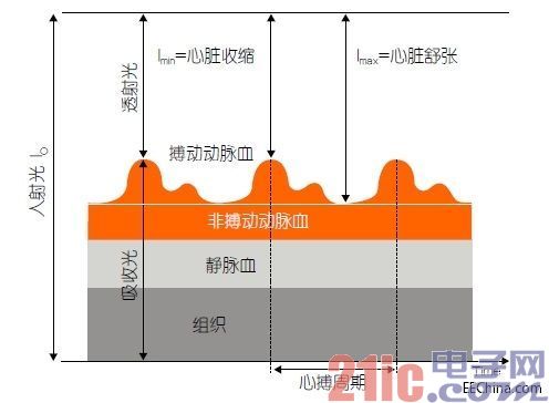
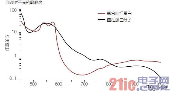
More and more people track their physical fitness through wearable widgets and the right application software. Optical sensors are suitable for measuring pulse rate and oxygen saturation. This technology is very mature in the medical field and can now be ported to consumer applications thanks to modern LED technology. This article refers to the address: http:// All of this can be counted from the bracelet that records the number of steps people walk. Now, a variety of motion trackers such as fitness bracelets and smart watches can also measure heart rate and other biological indicators or monitor sleep quality. Many people are very optimistic about new opportunities to track their fitness level, which has led to the increasingly popular "quantitative self" movement. Large companies such as Samsung, Apple and Google are entering the growing market with the right apps, smart watches and smartphones. Although pedometers use accelerometers, optical methods commonly used in the medical field for pulse and oximetry are entering the consumer market. In a hospital setting, most of the sensors are mounted in the ears or finger clips. In 2013, Mio Alpha's smartwatch became the first bracelet to measure the pulse rate on the wrist using an optical sensor – a significant improvement over the chest strap worn by athletes. No one wants to wear the chest strap all day. On the body. Smartphones can also be used to measure pulse rate on a finger. The first fitness bracelets are now on the market, and you can measure the oxygen saturation in your blood by simply placing your finger on the screen. This feature is very useful, for example, for people working at high altitudes, such as climbers, gliders and glider guides, and people with heart or lung disease. Optical measurement method The principle of sensor measurement of pulse rate and oxygen saturation is called optical plethysmography (PPG), in other words, optical measurement of blood flow changes in blood vessels. This approach takes advantage of this principle: the blood flow delivered in the arteries varies regularly with the heart pumping cycle. The heart rhythmically pumps blood (heart contraction) and blood draw (heart relaxation) at regular intervals. This means that more blood flows through the artery during the systole phase and less blood flow during the diastolic phase. By measuring changes in blood flow in a particular part of the body, the pulse rate can be obtained from the periodicity of the signal being measured. Blood flow is measured by the ability of hemoglobin in the blood to absorb light (Figure 1). The sensor consists of a light source and detector placed next to each other and placed directly on the skin for measurement. The emitted light penetrates into the skin, tissues and blood vessels and is absorbed, emitted and reflected. The intensity of the reflected light recorded by the detector will vary depending on the flow of blood flowing through the artery (Figure 2). The appropriate wavelength for this measurement depends on which part of the body is being measured. Green light provides the best results at the wrist, while red and infrared light is typically used for measurements at the fingertips. Figure 1: Principle of reflected light pulse measurement. Light from the sensor passes through the skin and tissue, a portion is absorbed, and a portion is reflected back to the detector. Because the blood flow in the artery changes with each beat of the heart, the amount of light absorbed and the signal strength received by the detector change. Green light can provide the best results at the wrist, while red and infrared light is generally used for finger measurements. Figure 2: Generation of detector signals in PPG measurements. Light (I0) that illuminates the skin is absorbed by venous or arterial blood or reflected back to the detector. The varying component of the signal corresponds to the arterial blood flow that changes in synchrony with the heartbeat. The period of change of this signal indicates the pulse rate. The ratio of the minimum and maximum detector signals (photocurrent Imin/Imax) provides the basis for determining blood oxygen saturation. If the absorption rate is measured by infrared light and red light, the oxygen saturation in the arterial blood can be determined. Known as a pulse oximeter, this method is the only non-invasive method for determining blood oxygen saturation. In other words, it is the only method that does not require blood collection. The pulse oximeter takes advantage of the fact that the oxygen concentration in the blood is different and the amount of light absorbed is also different. Oxygen is transported through the hemoglobin molecule (Hb) in the blood. When Hb binds to oxygen molecules, it forms oxyhemoglobin (HbO2), and its absorption properties also change (Fig. 3). The concentration of the two hemoglobin molecules (cHbO2 and cHb) in the blood indicates the oxygen saturation SpO2: SpO2 = cHbO2 / (cHbO2 + cHb). Figure 3: Hemoglobin in the blood - The light absorption properties of hemoglobin (Hb) change when combined with oxygen molecules (oxygenated hemoglobin or HbO2). Blood oxygen saturation can be determined by a PPG measurement method using red light and infrared light. The concentration of light absorbing material in the radiation medium is a function of the light absorption rate. In PPG measurements, there are two different wavelengths of light used to obtain reliable cHbO2 and cHb indications. Red light with a wavelength of 660 nm (nm) and infrared light with a wavelength of 940 nm are well suited for this application because the two hemoglobin molecules (Hb and HbO2) have the most different absorption properties for the two types of light (Figure 3). In order to determine the oxygen saturation in the artery, we need to check the absorption of the pulse signal component (Figure 2). Oxygen saturation (SpO2) can be expressed as a function of the ratio of the minimum to maximum detector signals (Imin and Imax) for the relevant wavelength conditions. It is the development of thin film chip technology that makes it possible to produce high efficiency LEDs with a narrow spectral bandwidth of about 30 nm. This technique also ensures higher system efficiency because the thin-film LED emits almost all of the light from the top, so essentially all of the light can be fully utilized. The design must also ensure that the wavelength remains stable throughout the measurement, although the chip will have a temperature rise during the measurement. In addition to the good thermal stability of LEDs, short pulses are also a good way to maintain wavelength stability. A pulse having a pulse length of less than 0.3 ms and a repetition rate of about 2 ms is preferable. The choice of wavelength depends on the measurement you want to make. For sensors worn on the wrist, green LEDs with a wavelength of about 530 nm are preferred; finger sensors typically use red (660 nm) or infrared (940 nm) light. LEDs are available in a variety of versions so they can be adapted to different designs and applications. It is sufficient to use one wavelength for the pulse sensor, and the measurement of blood oxygen saturation usually uses infrared and infrared light alternately. For detectors, key requirements include high linearity, excellent sensitivity, and good signal-to-noise ratio. Linearity is especially important when measuring blood oxygen values ​​because the absolute photocurrent values ​​Imin and Imax must be measured very accurately. Large area photodiodes with low dark currents are suitable, such as SFH 2400 or SFH 2430 with ambient light filter. Photodiodes also provide fast switching times and therefore correspond well to the required short LED pulses. Integrated sensors like the SFH 7050 are a compact solution. This multi-chip sensor consists of 3 LEDs and 1 photodiode designed for pulse and oximetry measurements in wearables and smartphones. The high-efficiency LED is made of thin-film technology, and the emitted light (green is 530nm, red is 660nm, infrared is 940nm) has a narrow bandwidth, so this sensor can support the pulse rate measurement at the wrist and the blood on the finger. Oxygen saturation measurement. The integrated light barrier is a good way to prevent light from crosstalking from the LED to the photodiode. Depending on the application, the three LEDs can be operated individually or in turn. Infrared LEDs in the SFH 7050 can also be used in combination with a receiver as a proximity sensor to automatically start or stop measurements when the sensor contacts or leaves the skin. Chipsets for PPG and pulse oximetry in medical applications are already available on the market and can be used to control LED and digital detector signals. TI's TI AFE 4403 is a good example of excellent digital resolution (22-bit). Sensor design As with all sensors, signal quality is one of the most important considerations when designing optical sensors for pulse rate and oxygen saturation measurements. The complete detector signal consists of a very large constant (DC) component and a small variable (AC) component, corresponding to pulsed arterial blood (Figure 2). For example, measurements on a light-skinned human wrist using an LED with a wavelength of 530 nm and a current of 8 mA will produce an AC/DC ratio of approximately 0.00035. For a dark-skinned person, this value is lower, but the measurement on the finger may be about 10 times higher. As a small part of the overall signal, this particularly small AC signal is a daunting challenge for the digitization process – especially when measuring blood oxygen values. In this case, the entire signal must be digitized so that the resolution of the AC component and its maximum and minimum values ​​(Imin and Imax) can be high. In actual use, this means that the overall signal requires a resolution of at least 16 bits. If you only need to measure the heart rate, then only the periodic variation of the signal is important, and the absolute value of the signal can be ignored. In this case, the constant component can be suppressed by a bandpass filter, and the remaining AC component is amplified to the AC/DC converter. The minimum and maximum values ​​of the photocurrent are required to achieve high resolution, and it is also important to measure against the skin. Typical sampling rates are between 25 Hz and 500 Hz per channel, and LED pulse lengths are in the range of 0.5 ms to 5 ms. Another factor is ambient light. Even if the sensor is properly placed on the skin, ambient light can reach the receiver because the infrared light can penetrate deep into the skin and diverge inside. Ambient light will have a negative impact on the signal-to-noise ratio and therefore need to be suppressed as much as possible, such as by good contact between the sensor and the body, or by a dedicated ambient light filter on the detector. This filter converts the detector's maximum sensitivity from the infrared spectrum to the visible spectrum, such as to 570 nm with the photodiode SFH 2430, making it ideal for green LEDs. One common method for eliminating ambient light effects in detector signals is to make two measurements with and without LEDs, and then take the difference between the two signals. A chipset like the TI AFE 4403 can also emit a suitable dark signal for this purpose. to sum up With the continued development of quantitative self-movement, optical sensors for medical applications are beginning to penetrate into the consumer electronics arena, especially fitness bracelets, smart watches and smartphones. This is due to the high efficiency LEDs with excellent spectral purity. Integrated optical sensors for measuring pulse rate and oxygen saturation represent the development of technology in this growing market.
32V DC Power Supply is applicable to electrical equipment testing sector. The output current for 1U unit and 2U unit is up to 50A and 200A respectively.
It also covers 12V DC Power Supply, have the features including high precision and resolution,high efficiency low ripple ,complete multiple protection (OVP/OCP/OPP/OTP/SCP), Master/Slave Series/Parallel modes up to 10 units, and multiple communication interfaces,which allow a more flexible and convenient use of the Programmable Power Supply.
Some features of the DC Power Supply as below:
32V DC Power Supply 32V DC Power Supply,Adjustable DC Power Supply,Switching Power Supply,Computer Power Supply APM Technologies (Dongguan) Co., Ltd , https://www.apmpowersupply.com